
SIMANNU Simulation Centre presents its homemade vascular access simulators that offer realistic and safe training, improving operators’ skills and reducing risks in clinical procedures. These do-it-yourself devices represent a cost-effective solution that lowers acquisition and maintenance costs compared to traditional commercial models. Their use makes it possible to extend advanced training to a larger number of healthcare professionals, democratising access to cutting-edge training technologies.
Importance of Precision in Vascular Procedures
On a cold November morning, an error during a routine insertion of a central venous catheter in a hospital turns a standard procedure into a complication to be managed. Inaccurate insertion can not only cause bleeding, more or less severe, but can also put the patient at risk of long-term complications, underlining a disturbing truth: precision in such procedures is not only desirable, but essential.
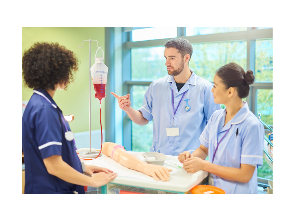
Today, vascular access simulators, advanced training tools that replicate the challenges of real-life medical procedures, emerge as essential in preventing such errors, offering healthcare professionals a safe and controlled environment to hone their skills before tackling real-life situations. In the field of ultrasound, for example, they represent a valuable support for both medical and nursing clinical learning manoeuvres, and should be considered as true safety and clinical risk reduction tools.
Barriers and Innovative Solutions: the SIMANNU experience
However, the adoption of these advanced training tools faces a significant obstacle: cost. Vascular access simulators represent a substantial investment, with prices that can vary enormously depending on the complexity and technology employed. This high cost can limit the availability of such training devices, especially in resource-limited settings.
A promising solution is emerging due to the growing adoption of in-house manufacturing: the do-it-yourself production of simulators. This approach offers numerous advantages, including significant cost reductions and the ability to customise simulators to meet specific training needs.
The trainer tasks that we present in this article are fully produced at the SIMANNU simulation centre in Nuoro, Italy, and are the result of several phases of development and experimentation, in order to respond more satisfactorily to participants’ training needs.
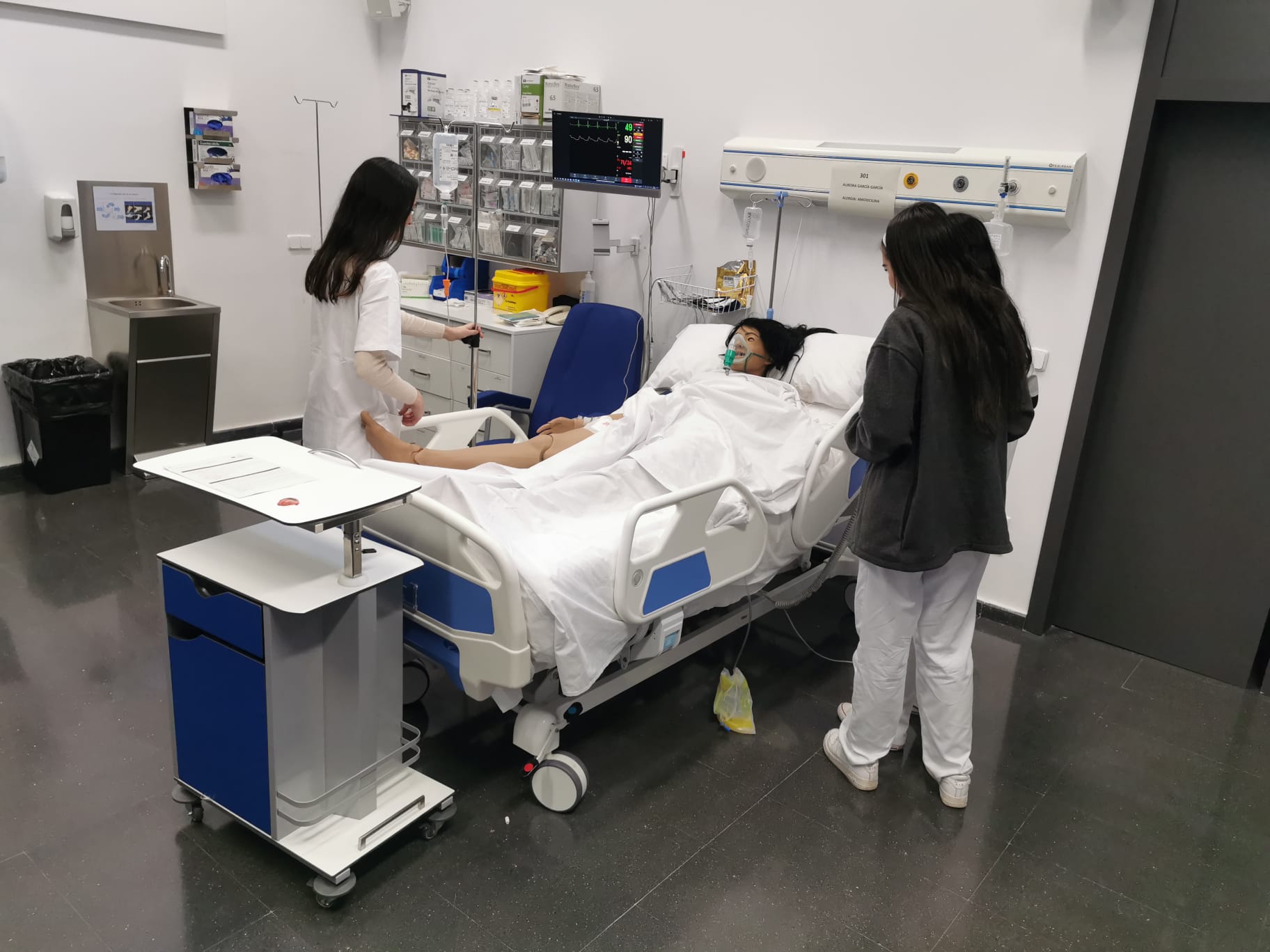
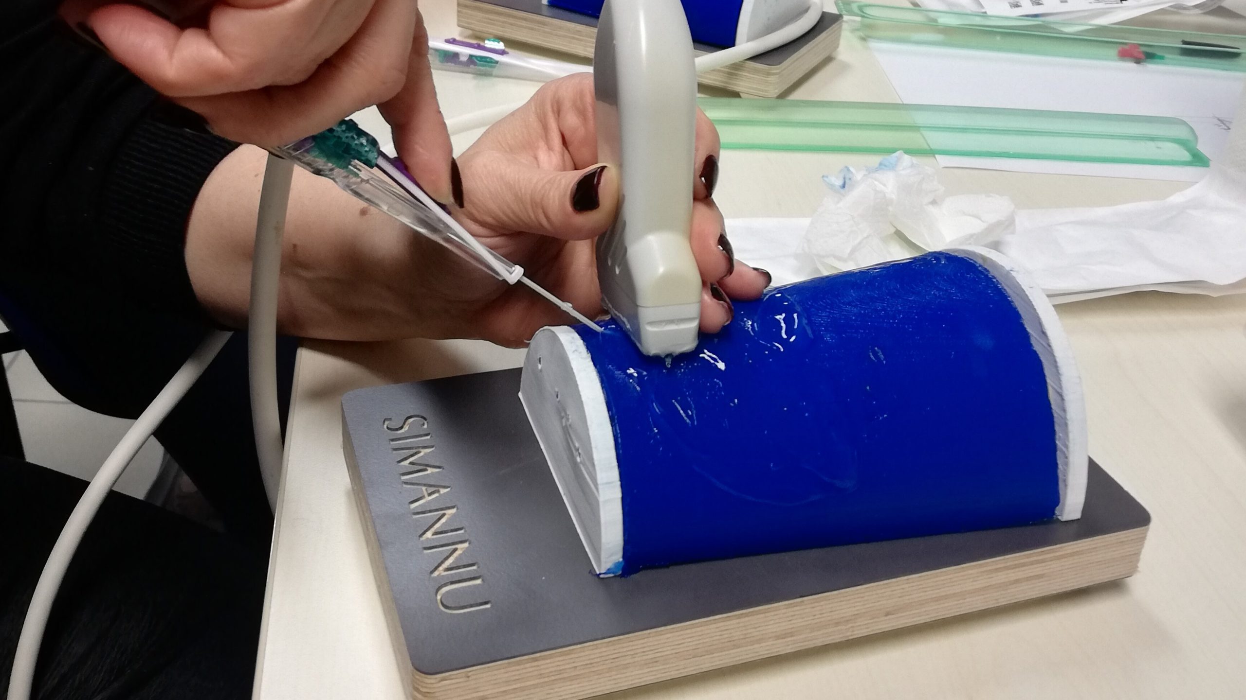
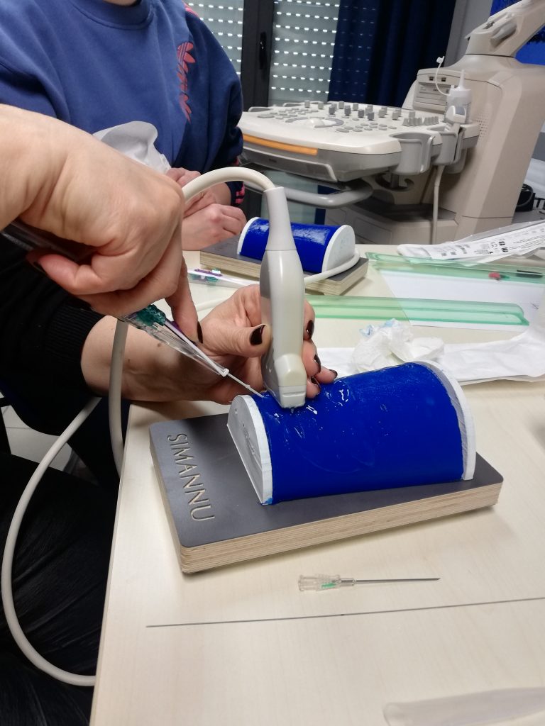
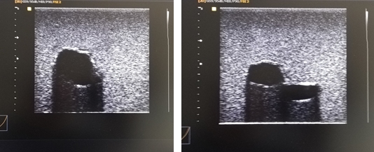
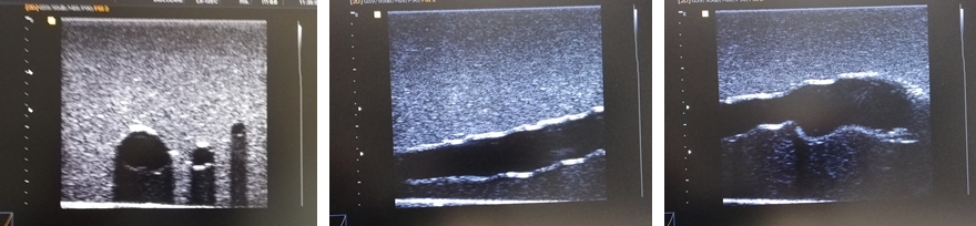
The tasks, the specifications of which we will provide below, make it possible to simulate venipuncture with a cannula needle, as well as with a syringe, up to the implantation of PICCs, Midlines and central venous catheters (CVCs) in an extremely faithful manner. Blood sampling, blood culture and haemogas analysis can also be simulated. The versatility of these devices makes it possible to train medical and nursing professionals, as well as medical and nursing school students, in various clinical practices. During training, participants can safely simulate the invasive procedure and realistically visualise the puncture of the vessel through the ultrasound screen, thus developing the specific skill (SimZone 1). In order to train technical and non-technical (human factors) skills in an integrated manner, an advanced version of the device is placed on a simulated patient, enabling the realisation of complex scenarios (SimZone 2).
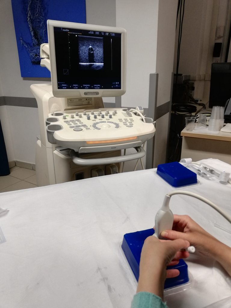
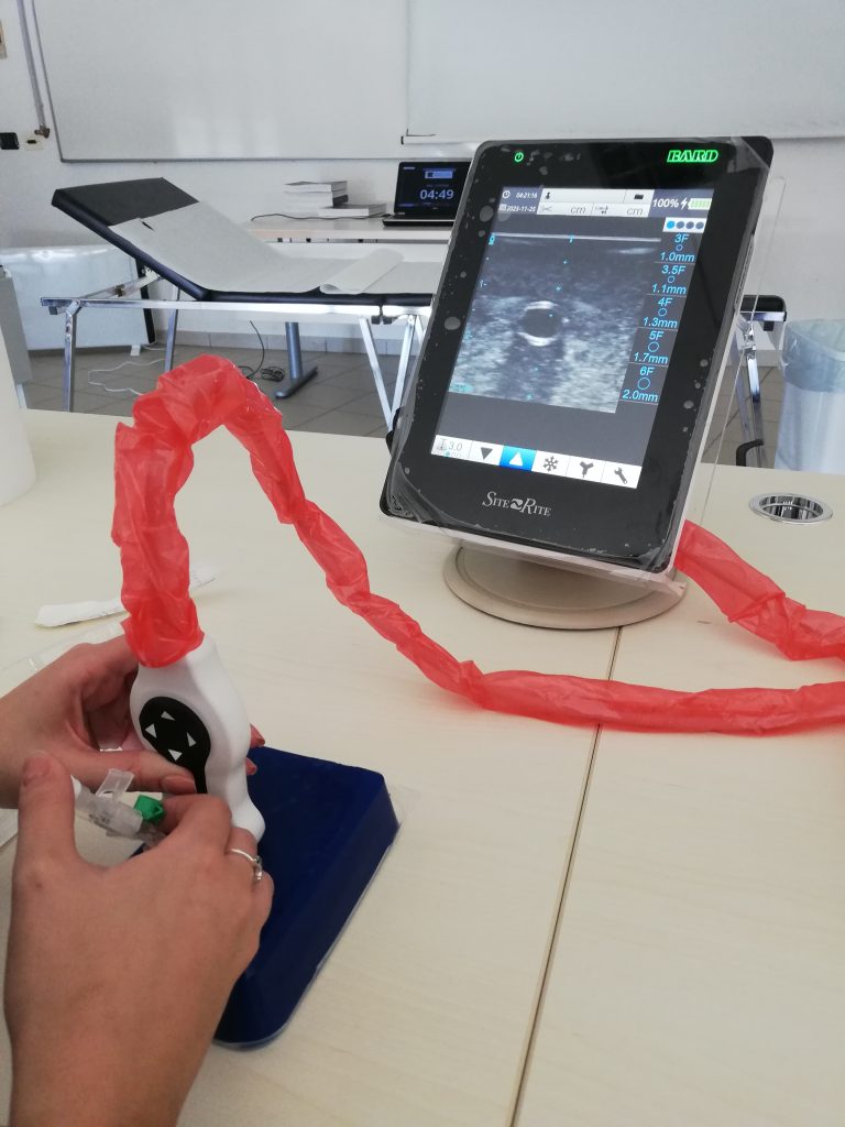
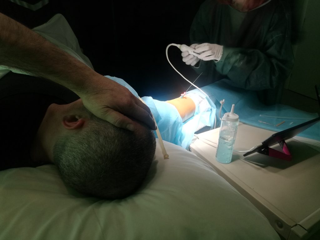
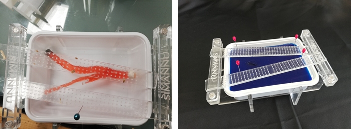
We developed two types of devices: one ‘square and flat’ and one ’rounded’. The first has proved to be valid for accessing the vessel, as it develops psychomotor coordination between the dominant hand using the needle and the other hand holding the ultrasound probe. The second, in addition to accustoming the operator to working on curved surfaces, such as the arm, is used both at skills stations and during advanced training and simulation scenarios, realistically reproducing the anatomical part and the vascular district where the insertion is made.
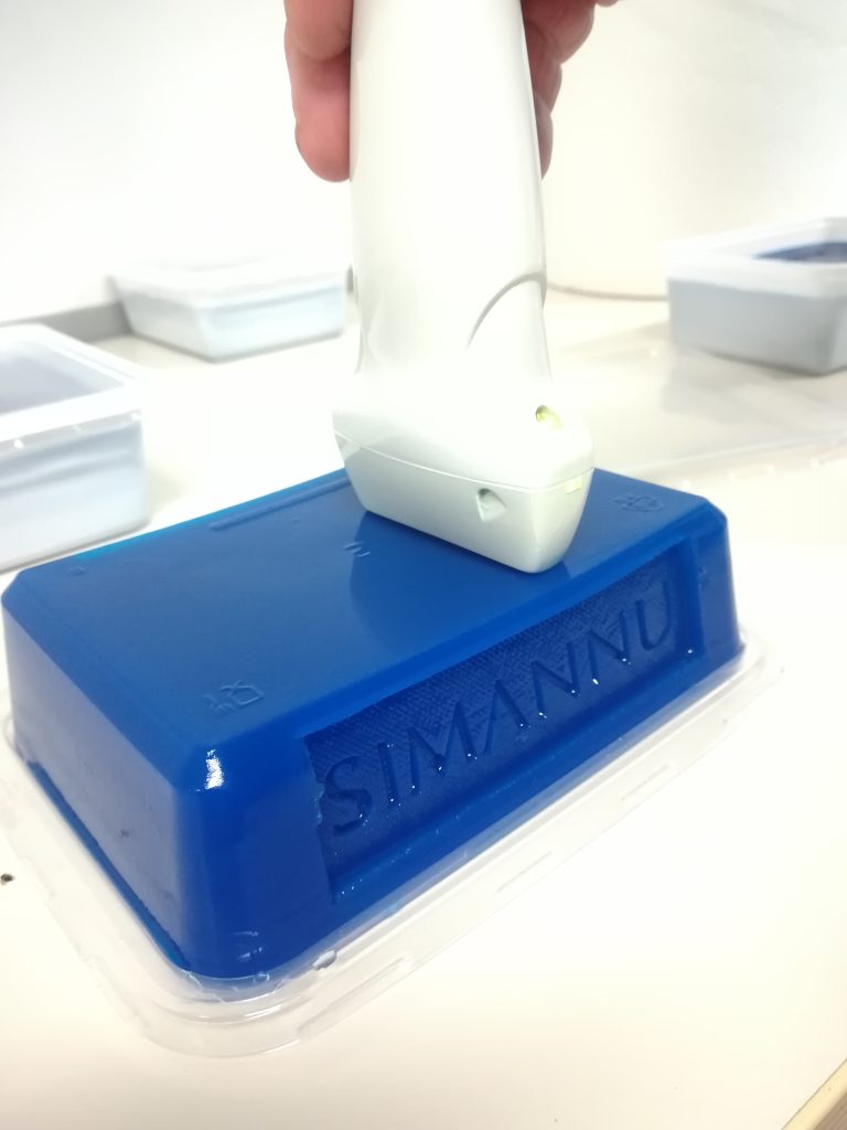
The main ingredient for making ultrasound PADs is AGAR powder, appropriately mixed with dyes and substances that retard physiological degradation.
In recent years, we have made several attempts with various products (ballistic gel, isinglass, gelatine powder), but Agar turned out to be the material with the best compromise between technical characteristics (acoustic parameters, attenuation coefficient and acoustic impedance) and cost-effectiveness. In-depth studies and a research study are currently underway to produce products that meet the diverse requirements of this training sector.
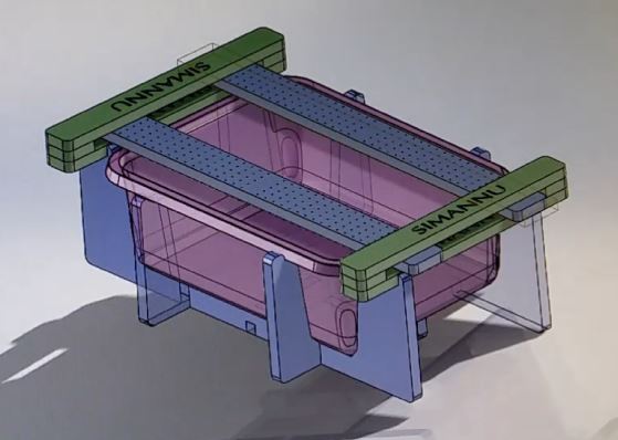
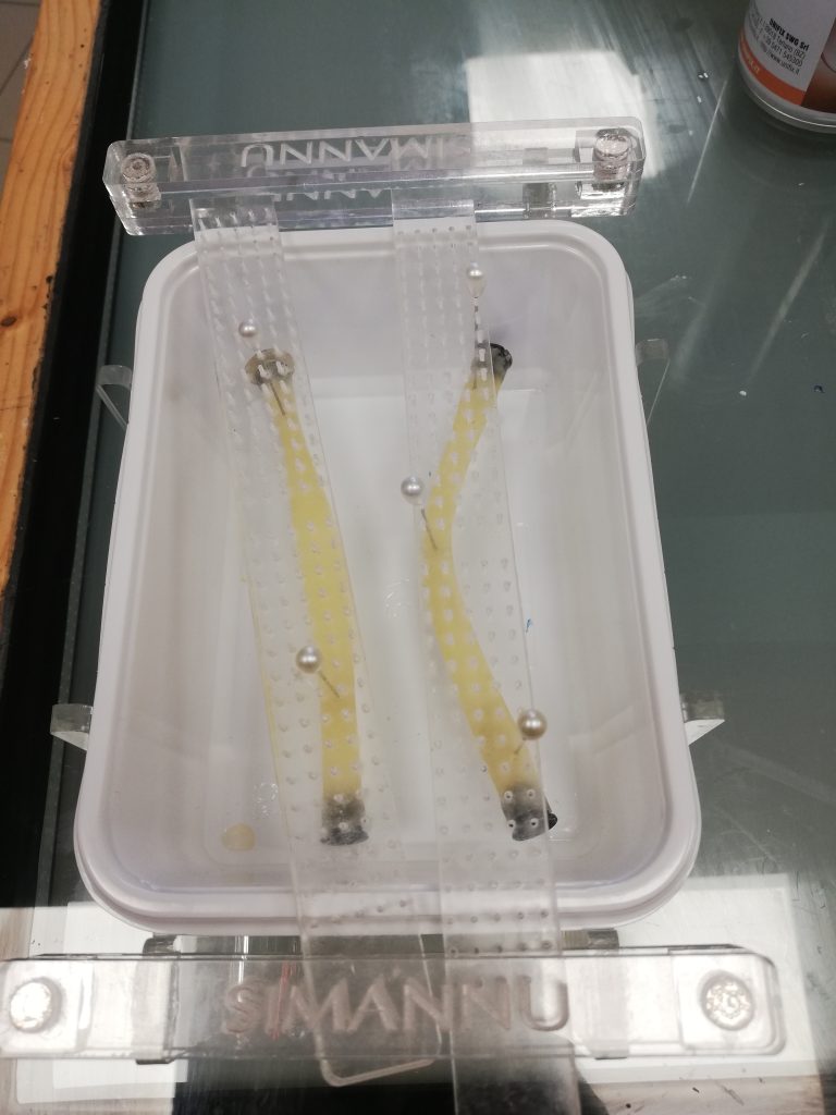
In order to realise PADs with different shapes, we have designed and manufactured the supports that will hold the Agar. In particular, we developed a support for the ‘flat’ PAD, made of precision laser-cut plexiglass. The peculiarity of this support lies in its ability to support and orient/curve the tubes simulating the veins. This feature allows each participant to have a unique PAD, replicating in a certain way the anatomical variability of real patients. In order to obtain a realistic image, the tubes are filled with a liquid, even just coloured water.
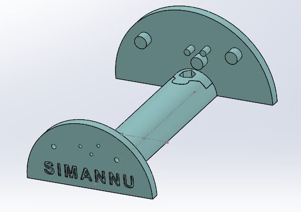
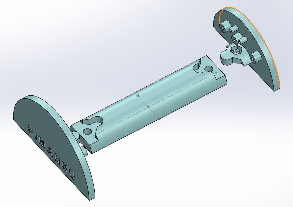
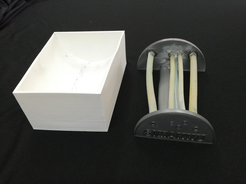
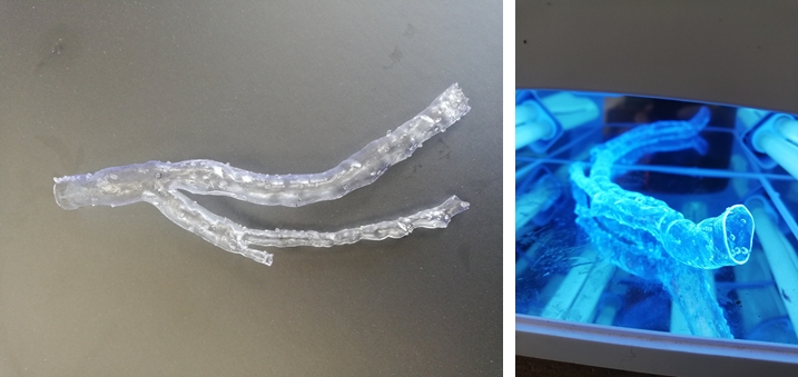
For the ’round’ PAD, we adopted a 3D printing approach. Two supports were made: one dedicated to the PAD itself, with a detailed vessel system including the cephalic vein, basilic vein and brachial veins and arteries; and a global support to contain the PAD during the drying periods.
Continuous Innovation and Development
With the aim of developing more and more durable and ultrasound-compatible products, we are working on creating PADs with an increasingly true-to-life vessel system. Today, we are also able to extract the anatomical part of interest from a clinical image in DICOM format (such as a CT scan or MRI). Using ‘segmentation’ software, we can isolate the part of interest and subsequently 3D print it.
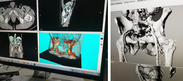
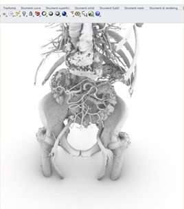
A practical example of this process is shown in the pictures, where we performed the segmentation and export of a complete abdominal aorta.
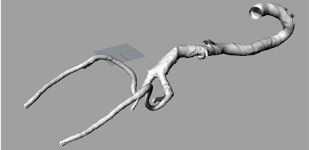

In the specific context of vascular surgery, we extracted a segment of the femoral aorta for the creation of a specialised training PAD.
All this was possible because in recent years a prototyping laboratory has been set up within Simannu for the very purpose of creating simulators and task trainers.
It might seem demanding in terms of cost, time and the effort required, but in reality they are simple and inexpensive processes.
The creation starts with the 3D design of models. In our case, SolidWorks 3D modelling software was used. The cost of the software is high, but there are also free alternatives on the market with free licences for students and teachers.
The next step is the printing of the models. In our case, a support for the casting and a support for the PAD were printed. The printer used is an Ender-3, an inexpensive and easy-to-use machine (approx. €170).
PLA, the most common, cheap and easy-to-use material, was used as the printing material.
For our models it took about 250g of PLA (a 1kg reel costs about 17€) and 9 hours of printing.
The material used for the ultrasound PAD is AGAR, a cheap and easily available food thickener. 100g of agar costs about 10€.
On the other hand, CT and MRI prints were made with the FormLab 3L, a resin printer.
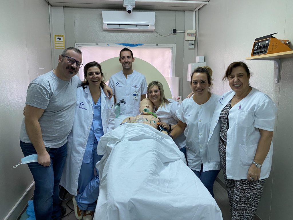
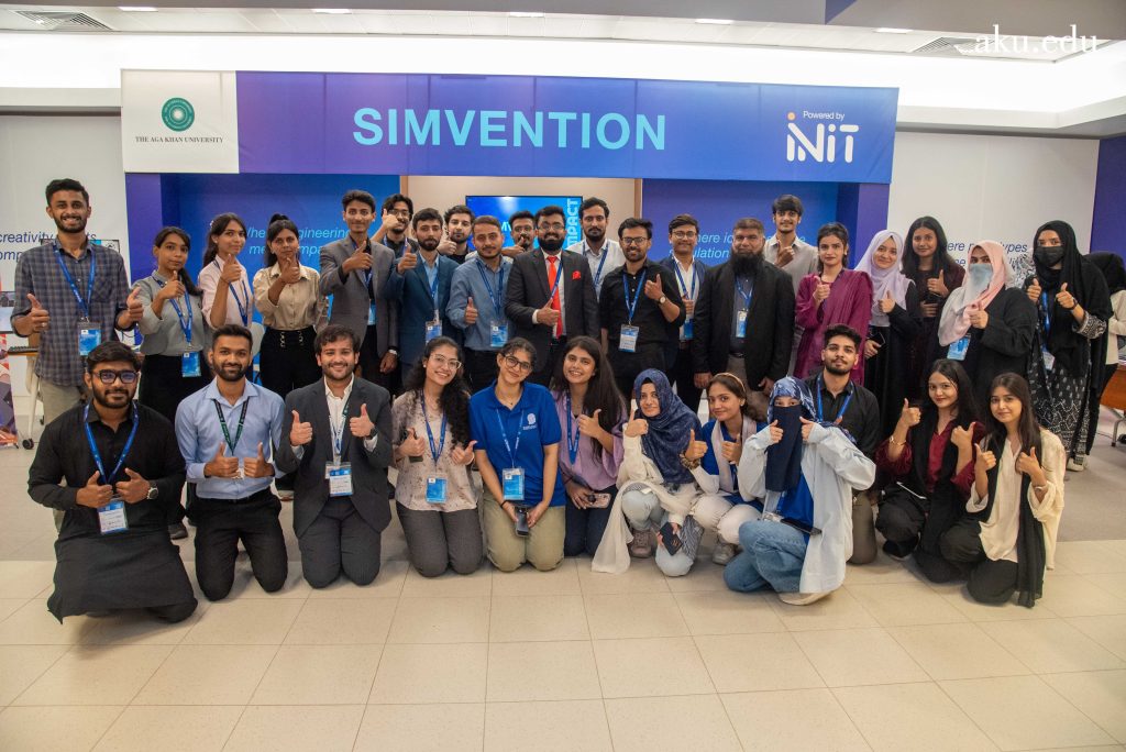
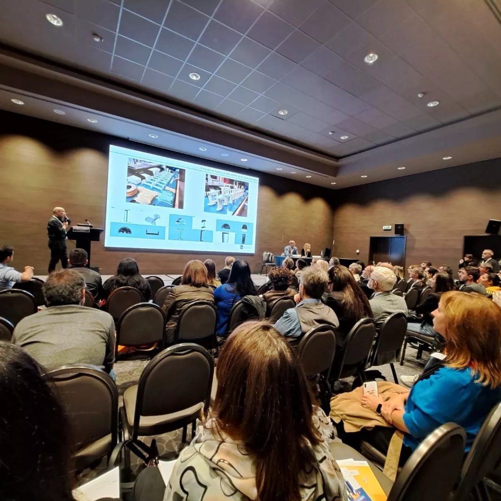
The results of our RD activities were presented at the 4th IVAS (Italian Vascular Access Society) National Congress held in Rome on 10 and 11 November 2023, as part of the session ‘New technologies for simulators for training and implantation training of vascular devices‘.
Conclusions
The evolution of simulators for the ultrasound-guided implantation of vascular devices highlights a growing need for innovation in the training of healthcare personnel. The task trainers implemented in our Simulation Centre in Nuoro have proven to be safe and realistic for training, improving the simulation of invasive procedures and reinforcing the specific skills of operators. Moreover, the diversity of manageable tasks, including venipuncture, vascular device installation and other clinical practices, confirms the versatility of these tools.
READ ALSO